3.1 SAS ?
3.1.1. Scattering profile ?
SAS data used in this integrative model was obtained from 5 deposited SASBDB entry (entries). Scattering data from solutions of biological macromolecules are presented as both log I(q) vs. q and log I(q) vs. log (q). The I(q) is the scattering intensity (preferably on an absolute scale in cm-1, but arbitrary units are accepted) and q is the modulus of the scattering vector (nm-1 or Å-1).
3.1.2. Key experimental estimates ?
Molecular weight (MW) estimates from experiments and analysis: Theoretical MW can be compared to SAS-derived values using the forward scatter (I(0)) and the known concentration and partial specific volume of the scattering particle, or as estimated from the Porod volume and partial specific volume (Trewhella et al., 2017, Trewhella et al., 2023).
| SASDB ID | Chemical composition MW | Standard MW | Porod Volume/MW |
|---|---|---|---|
|
SASDBV9
|
12.6 kDa
|
12.2 kDa
|
Not available
|
|
SASDBW9
|
24.1 kDa
|
25.2 kDa
|
Not available
|
|
SASDBZ9
|
49.4 kDa
|
48.3 kDa
|
Not available
|
|
SASDBX9
|
12.5 kDa
|
14.7 kDa
|
Not available
|
|
SASDBY9
|
25.9 kDa
|
25.2 kDa
|
Not available
|
Volume estimates from experiments and analysis: estimated volume can be compared to Porod volume obtained from scattering profiles.
| SASDB ID | Estimated Volume | Porod Volume | Specific Volume | Sample Contrast | Sample Concentration |
|---|---|---|---|---|---|
|
SASDBV9
|
Not available
|
17.94 nm³
|
Not available
|
Not available
|
Not available
|
|
SASDBW9
|
Not available
|
22.50 nm³
|
Not available
|
Not available
|
Not available
|
|
SASDBZ9
|
Not available
|
66.59 nm³
|
Not available
|
Not available
|
Not available
|
|
SASDBX9
|
Not available
|
56.68 nm³
|
Not available
|
Not available
|
Not available
|
|
SASDBY9
|
Not available
|
27.97 nm³
|
Not available
|
Not available
|
Not available
|
3.1.3. Flexibility analysis ?
In a Porod-Debye plot, a clear plateau is observed for globular (partial or fully folded) domains, whereas flexible-modular, fully unfolded domains or extended/stiff rodshaped domains lack a discernible plateau (Rambo and Tainer 2013). A bell-shaped Kratky plot (q²I(q) vs. q) with a well-defined maximum is observed for compact/folded structures. For partially flexible/modular or extended structures the Kratky plot can show multiple maxima and/or an increase in intensity at higher q-values depending on the degree of flexibility and extension. Fully intrinsically disordered structures yield a Kratky plot that systematically increases with increasing q values and will be near linear for highly extended molecules. The dimensionless Kratky plot ((qRg)²I(q) vs. qRg) is useful for quantifying differences in shape and foldedness among scattering objects of different sizes (Trewhella et. al., 2023).
Flexibility analysis for SASDBV9.
Flexibility analysis for SASDBW9.
Flexibility analysis for SASDBZ9.
Flexibility analysis for SASDBX9.
Flexibility analysis for SASDBY9.
3.1.4. Pair-distance distribution analysis ?
The the atom-pair distance distribution function (PDDF) or P(r) represents the distribution of distances between all pairs of atoms within the particle weighted by the respective scattering contrasts (Moore, 1980). The second moment of P(r) yields the radius of gyration (Rg), which is a measure of the overall size and shape of a macromolecule (i.e. the spatial distribution of volume elements). A protein with a smaller Rg is more compact than a protein with a larger Rg, provided both have the same molecular weight.
| SASDB ID | Software used | Dmax | Dmax error | Rg | Rg error |
|---|---|---|---|---|---|
|
SASDBV9
|
GNOM 4.5a
|
6.660 nm
|
Not available
|
1.824 nm
|
0.006 nm
|
|
SASDBW9
|
GNOM 4.5a
|
9.370 nm
|
Not available
|
2.787 nm
|
0.007 nm
|
|
SASDBZ9
|
GNOM 4.5a
|
15.430 nm
|
Not available
|
4.629 nm
|
0.011 nm
|
|
SASDBX9
|
GNOM 4.5a
|
7.930 nm
|
Not available
|
2.636 nm
|
0.008 nm
|
|
SASDBY9
|
GNOM 4.5a
|
10.450 nm
|
Not available
|
2.976 nm
|
0.005 nm
|
P(r) for SASDBV9: The value of P(r) should be zero beyond r=Dmax.
P(r) for SASDBW9: The value of P(r) should be zero beyond r=Dmax.
P(r) for SASDBZ9: The value of P(r) should be zero beyond r=Dmax.
P(r) for SASDBX9: The value of P(r) should be zero beyond r=Dmax.
P(r) for SASDBY9: The value of P(r) should be zero beyond r=Dmax.
3.1.5. Guinier analysis ?
The linearity of the Guinier plot (ln(q) vs. q²) at very-low angle (qRg < 1.3) is a sensitive indicator of the quality of the sample in relation to its homogeneity; a linear Guinier plot is a necessary but not sufficient demonstration that a solution contains monodisperse particles of the same size. Deviations from linearity can point to strong interference effects from particle attraction or repulsion, polydispersity of the samples, or improper background subtraction (Feigin et al., 2013). Residual difference plots and Pearson correlation coefficient determination (R²) are measures to assess quality of the linear fit to the Guinier region. A perfect fit has an R² value of 1. Residual values should be equally and randomly spaced around the horizontal axis with no evident systematic upward or downward curvature. Agreement between the P(r) and Guinier-determined Rg is a good measure of the self-consistency of the SAS profile.
| SASDB ID | Rg | Rg error | MW | MW error |
|---|---|---|---|---|
|
SASDBV9
|
1.77 nm
|
0.05 nm
|
12.2 kDa
|
Not available
|
|
SASDBW9
|
2.71 nm
|
0.06 nm
|
25.2 kDa
|
Not available
|
|
SASDBZ9
|
4.34 nm
|
0.17 nm
|
48.3 kDa
|
Not available
|
|
SASDBX9
|
2.78 nm
|
0.18 nm
|
14.7 kDa
|
Not available
|
|
SASDBY9
|
2.95 nm
|
0.11 nm
|
25.2 kDa
|
Not available
|
The linearity of the Guinier plot is a sensitive indicator of the quality of the experimental SAS data; a linear Guinier plot is a necessary but not sufficient demonstration that a solution contains monodisperse particles of the same size. Deviations from linearity usually point to strong interference effects, polydispersity of the samples or improper background subtraction. Residual value plot and coefficient of determination (R2) are measures to assess linear fit to the data. A perfect fit has an R2 value of 1. Residual values should be equally and randomly spaced around the horizontal axis.
3.2. Crosslinking-MS
At the moment, data validation is only available for crosslinking-MS data deposited as a fully compliant dataset in the PRIDE Crosslinking database. Correspondence between crosslinking-MS and entry entities is established using pyHMMER. Only residue pairs that passed the reported threshold are used for the analysis. The values in the report have to be interpreted in the context of the experiment (i.e. only a minor fraction of in-situ or in-vivo dataset can be used for modeling).
Crosslinking-MS dataset is not available in the PRIDE Crosslinking database.
3.3. 3DEM ?
This section describes quality of the 3DEM datasets
EMD-7321
3.3.1. Experimental information ?
| EM reconstruction method: | SUBTOMOGRAM AVERAGING | |||
| Resolution: | 28.00 Å | |||
| Recommended level: | 0.015 | |||
| Estimated volume: | 214542.20 nm³ | |||
| Specimen preparation: |
|
|||
| Map-only validation report: | wwPDB validation report |
3.3.2. Map visualisation ?
This section contains visualisations of the EMDB entry EMD-7321. These allow visual inspection of the internal detail of the map and identification of artifacts. Images derived from a raw map, generated by summing the deposited half-maps, are presented below the corresponding image components of the primary map to allow further visual inspection and comparison with those of the primary map.
3.3.2.1. Orthogonal projections ?
Primary map

|

|

|
| X | Y | Z |
The images above show the map projected in three orthogonal directions.
3.3.2.2. Central slices ?
Primary map

|

|

|
| X Index: 150 | Y Index: 150 | Z Index: 150 |
The images above show central slices of the map in three orthogonal directions.
3.3.2.3. Largest variance slices ?
Primary map

|

|

|
| X Index: 144 | Y Index: 156 | Z Index: 154 |
The images above show the largest variance slices of the map in three orthogonal directions.
3.3.2.4 Orthogonal standard-deviation projections (false-color) ?
Primary map

|

|

|
| X | Y | Z |
The images above show the map standard deviation projections with false color in three orthogonal directions. Minimum values are shown in green, max in blue, and dark to light orange shades represent small to large values respectively.
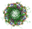
3.3.2.5. Orthogonal surface views ?
Primary map

|

|

|
| X | Y | Z |
The images above show the 3D surface view of the map at the recommended contour level 0.015 . These images, in conjunction with the slice images, may facilitate assessment of whether an appropriate contour level has been provided.
3.3.3. Map analysis ?
This section contains the results of statistical analysis of the map.3.3.3.1. Map-value distribution ?
The map-value distribution is plotted in 128 intervals along the x-axis. The y-axis is logarithmic. A spike in this graph at zero usually indicates that the volume has been masked.
3.3.3.2. Volume estimate ?
The volume at the recommended contour level is 214542.20 nm³.
The volume estimate graph shows how the enclosed volume varies with the contour level. The recommended contour level is shown as a vertical line and the intersection between the line and the curve gives the volume of the enclosed surface at the given level.
3.3.3.3. Rotationally averaged power spectrum ?
*Reported resolution corresponds to spatial frequency of 0.036 Å⁻¹
3.3.4. Fourier-Shell correlation ?
3.3.4.2. Resolution estimates ?
| Resolution estimate (Å) | Estimation criterion (FSC cut-off) | ||
|---|---|---|---|
| 0.143 | 0.5 | Half-bit | |
| Reported by author | 28.00 | - | - |
Author-provided FSC curve is not available.